  Female Anatomy The Female Human Being is made up of many parts and many systems, physical, mental and social. All of these change over her entire life!! As an Introduction to Women and Homoeopathy, it is vital that we know what makes up a WOMAN 
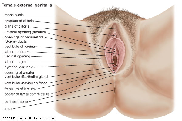
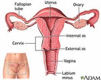
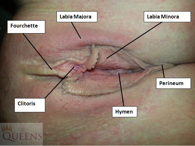
Anatomy of the Female Reproductive System 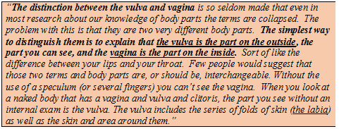 Vulva: The vulva is the external genitalia in the female reproductive system. Broadly the anatomical structures of vulva include:
 Vagina: The vagina is the internal muscular and tubular part of the female genital tract, which in humans extends from the vulva to the cervix. The outer vaginal opening may be partly covered by a membrane called the hymen. At the deep end, the cervix (neck of the uterus) bulges into the vagina. The vagina allows for, and channels, menstrual flow, which occurs periodically as part of the menstrual cycle. With regard to sexual activity, vaginal moisture is increased during sexual arousal for both human females and other female mammals. This is by way of vaginal lubrication, which reduces friction and allows for smoother penetration of the vagina during sexual activity. The texture of the vaginal walls creates friction for the penis during sexual intercourse and stimulates it toward ejaculation, enabling fertilization. Uterus: The uterus is a hollow, thick-walled, muscular organ situated deeply in the pelvic cavity between the bladder and rectum. Into its upper part the uterine tubes open, one on either side, while below, its cavity communicates with that of the vagina. It is divisible into two portions. On the surface, about midway between the apex and base, is a slight constriction, known as the isthmus, and corresponding to this in the interior is a narrowing of the uterine cavity, the internal orifice of the uterus. The portion above the isthmus is termed the body, and that below, the cervix. The part of the body which lies above a plane passing through the points of entrance of the uterine tubes is known as the fundus. The anatomy of the uterus consists of the following 3 tissue layers:
 Fallopian tubes: They are described in 4 parts (lateral to medial):
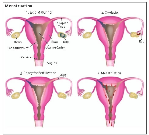
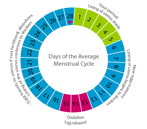
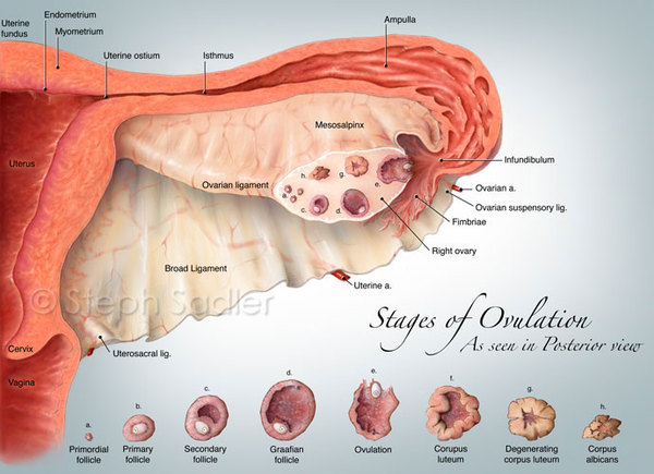
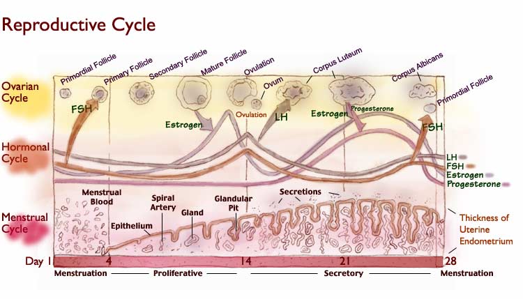
The main function of the uterine tubes is to assist in the transfer and transport of the ovum from the ovary, to the uterus. 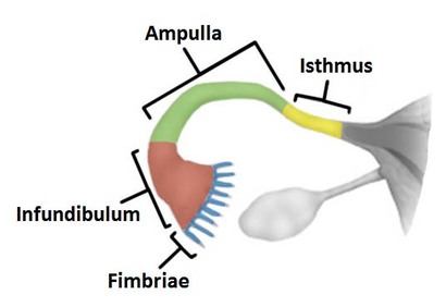 Ovary: The primary female reproductive organs, or gonads, are the two ovaries. Each ovary is a solid, ovoid structure about the size and shape of an almond. The ovaries are located in shallow depressions, called ovarian fossae, one on each side of the uterus, in the lateral walls of the pelvic cavity. They are held loosely in place by peritoneal ligaments. The ovaries are covered on the outside by a layer of simple cuboidal epithelium called germinal (ovarian) epithelium. This is actually the visceral peritoneum that envelops the ovaries. Underneath this layer there is a dense connective tissue capsule, the tunica albuginea. The substance of the ovaries is distinctly divided into an outer cortex and an inner medulla. The cortex appears denser and more granular due to the presence of numerous ovarian follicles in various stages of development. Each of the follicles contains an oocyte, a female germ cell. The medulla is loose connective tissue with abundant blood vessels, lymphatic vessels, and nerve fibers. 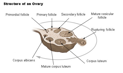 Physiology of ovarian and menstrual cycle: The hormonal cycle facilitates maturation and rupture of the ovarian follicle resulting in the release of an ovum (the female reproductive or germ cell). Each month a series of changes take place which prepares the uterus for pregnancy. The menstrual cycle is described below:
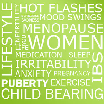
 Menopause Menopause, the end of menstruation, occurs between the ages of 45 and 55 (with the average age of 51.3). An entirely normal developmental and physiological process, it can be accompanied by symptoms including hot flashes, fatigue, moodiness, insomnia, decreased libido and sexual response, changes in memory, weight gain, and vaginal dryness. Until cessation of ovarian function is confirmed through a blood test, and/or one year of no menses, women may continue to ovulate and therefore require contraception to prevent unintended pregnancy. 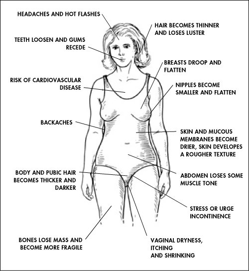
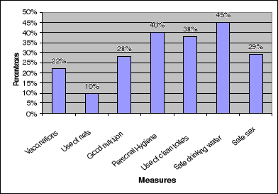
Risk Factors That Can Keep You from Living a Healthy Life Some diseases cannot be avoided. However, there are things all women can do to improve their health. Making healthy choices can help prevent or delay many of the common chronic diseases seen in women. Major threats to women’s health include:
How can you manage to lead to healthy life?
Effects of smoking 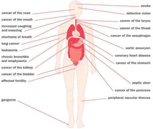

 Biblography:http://www.columbia.edu/itc/hs/pubhealth/modules/reproductiveHealth/anatomy.html
http://www.webmd.com/drugs/index-drugs.aspx http://emedicine.medscape.com/article/169450-overview https://en.wikipedia.org/wiki/Menstrual_cycle http://www.mayoclinic.org/healthy-lifestyle/womens-health/in-depth/menstrual-cycle/art-20047186?pg=1
0 Comments
|
CategoriesAuthorA convinced believer, an eternal student and a fervent practitioner of Homeopathy and Alternative Medicines, I am awestruck by the real-life miracles these sciences deliver every day! My goal is to help people with my knowledge, experience and willingness to learn and adapt! |


 RSS Feed
